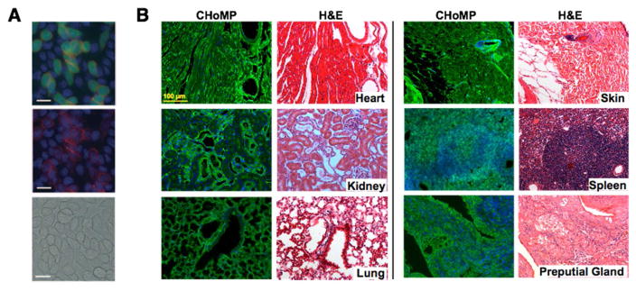
Figure 6. Chemoenzymatic glycan detection applications.
(A) Imaging of cell-surface glycans with LacNAc. Lec2 cells stained with chloromethyl fluorescein diacetate (CMFDA, green) and unstained Lec8 cells were mixed at a 1:5 ratio and were cultured for 3 days. The cells were treated with 500 mM GDP-FucAz and 30 mU α1–3 FucT for 15 min, then labeled with 20 mM DIFO-647 for 25 min. Top: Alexa Fluor 647 image merged with fluorescein (green) and Hoechst 33342 stain (blue); middle: Alexa Fluor 647 image merged with the Hoechst 33342 image; bottom: the bright-field image. Scale bars: 20 mm. Reprinted from 40 with permission from Wiley-VHC Verlag & Co © 2011. (B) CHoMP LacNAc labeling method applied to 5 μm, FFPE mouse heart, kidney, lung, skin, spleen, and preputial gland tissues, next to serial sections of the same tissues stained with H&E (Hematoxylin and eosin). Green: LacNAc staining; Blue: DAPI nuclear staining. Reprinted from 47 with permission from Wiley-VHC Verlag & Co © 2014.

