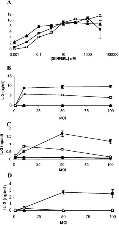
FIG. 4.
Antigen presentation from recombinant mycobacteria by dendritic cells. DC2.4 (□), BMDC (▪), and splenic dendritic cells (▴) were pulsed with increasing concentrations of SIINFEKL peptide for 2 h, washed, and then cultured with B3Z for 16 h. IL-2 production in supernatants was measured (A). Various dendritic cells were infected with increasing amounts of M. bovis BCG (▴), M. bovis BCG-OVApet (▪), M. smegmatis (▴), or M. smegmatis-OVApet (□) and cultured with the B3Z hybridoma for 16 h. Presentation of SIINFEKL on H2-Kb was measured by IL-2 secretion. The dendritic cell line DC2.4 (B), bone marrow-derived dendritic cells (C), and CD11c+ splenic dendritic cells (D) are shown.

