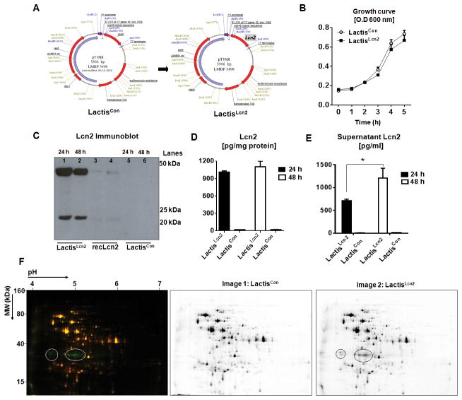
Figure 1. Schematic representation and characterization of Lcn2 expressing L. lactis.
(A) Murine Lcn2 gene was cloned into pT1NX plasmid at BamH1/Spe1 sites downstream of P1 promoter and USP45 signal sequence to allow for constitutive expression and secretion, respectively. (B) Line graphs represent the growth curve for the recombinant LactisLcn2 and LactisCon cultured in M17 media supplemented with erythromycin. LactisLcn2 and LactisCon were cultured for 24 and 48 h, and Lcn2 levels were assayed in the cell lysates by (C) immunoblotting and by (D) ELISA, and (E) culture supernatant by ELISA. Mouse recombinant (rec)-Lcn2 was used as a positive control for the Lcn2 immunoblot. (F) Composite overlay representative 2D-DIGE image (left) and individual images (right) showing protein accumulation in LactisCon (Cy5, red fluorescence) and LactisLcn2 (Cy3, green fluorescence) lysates. Green spots represent proteins up-regulated in LactisLcn2 and yellow spots represent proteins that are not deregulated. Circles indicate the protein bands predicted to be Lcn2. All experiments were performed in triplicates and are representative of three independent experiments. Results are expressed as mean ± SEM; *p<0.05.

