Figure 4.
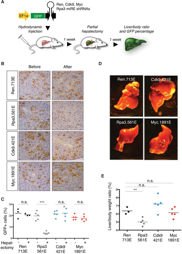
Liver regeneration for assessing toxicity of Cdk9 suppression in vivo. (A) Schematic representation of the liver regeneration. miR-E short-hairpin RNA (shRNA) transposon vectors targeting control Renilla luciferase, Cdk9, Myc, or Rpa3 were injected together with CMV-SB13 transposase by hydrodynamic tail vein injection into female C57BL/6 mice (6–8 wk of age) from Harlan Laboratories as previously described (Huang et al. 2014). Partial (2/3) hepatectomy (Taub 2004) was performed after 1 wk. Before being anesthetized with isoflurane, the animals received one subcutaneous dose of buprenorphine (0.5 mg/kg) and meloxicam (2 mg/kg). The median lobe, with its two central portions together with the left lateral lobe were mobilized, looped with a 4-0 vicryl ligation (Ethicon), and two-thirds of the liver volume were removed. Liver/body ratio and green fluorescent protein (GFP) percentage were examined after 2 wk. (B) Representative histological analysis of livers stained for GFP. Scale bar, 50 µm. (C) Cdk9 or Myc suppression does not show significant impact on the percentage of GFP+ cells before and after partial hepatectomy. (D) Representative images of livers injected with the indicated shRNAs after partial hepatectomy. (E) Cdk9 or Myc suppression does not show significant impact on liver/body ratio compared with Ren.713E (neutral control shRNA). Rpa3.561E is used as a positive control shRNA. All mouse experiments were approved by the Memorial Sloan Kettering Cancer Center (MSKCC) Animal Care and Use Committee (protocol no. 11-06-011). Statistical significance was calculated by two-tailed Student’s t-test (Prism 6 software). Significance values are *, P < 0.05; **, P < 0.01; and ***, P < 0.001.

