Fig. 3.

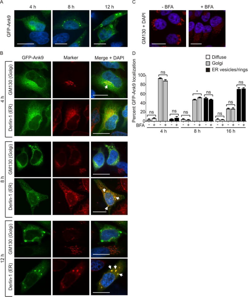
Ectopically expressed Ank9 exhibits temporal localization phenotypes that are consistent with Golgi-to-ER retrograde trafficking. Serum starved HeLa cells were transfected to express GFP-Ank9 (A and B). At 4, 8, or 12 h post transfection, the cells were fixed and examined by confocal microscopy (A–C). In some cases, fixed cells were screened with antibody against the cis-Golgi marker, GM130, or the ER protein, derlin-1 prior to confocal microscopic analyses (B). Representative fluorescence images of cells viewed for GFP; GM130 or derlin-1; and merged images plus DAPI are presented. White arrows in B denote representative points of GFP and organelle marker signal colocalization. (A) GFP-Ank9 exhibits subcellular localization patterns reminiscent of the Golgi at 4 h and destabilized ER at 8 and 12 h. (B) Immunofluorescent labeling confirms that GFP-Ank9 localizes to the Golgi at 4 h and subsequently traffics to the ER at 8 h and 12 h. Destabilization of GFP-Ank9-positive Golgi and ER is apparent at the 8 and 12 h time points. Representative cells exhibiting GFP-Ank9 Golgi like (Golgi) and ER-like (ER) subcellular localization patterns are denoted. (C and D) GFP-Ank9 does not require an intact Golgi to localize to the Golgi and ER. HeLa cells were treated with brefeldin A (BFA) or vehicle control. At 4 h, the cells were immunolabeled with GM130 antibody, stained with DAPI, and visualized using confocal microscopy to confirm that BFA destabilizes the Golgi (C). (D) Serum starved HeLa cells were treated with BFA- or vehicle control just prior to transfection with GFP-Ank9 plasmid. The cells were examined for GFP-Ank9 subcellular localization at 4, 8, and 16 h. Data are presented as the percentage of cells in which GFP-Ank9 signal was diffuse in the cytosol (Diffuse), localized to and disrupted the Golgi (Golgi), or localized to and altered the morphology of the ER to yield vesicular or ring-like structures (ER vesicles/rings). Results are representative of at least two separate experiments performed in triplicate, each yielding similar results. Scale bars, 20 μm. ns, not significant.
