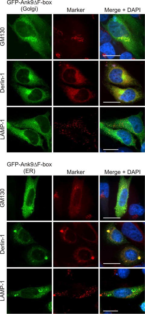
Fig. 4.

GFP-Ank9 localization to and perturbation of Golgi and ER morphology is F-box-independent. HeLa cells expressing GFP-Ank9ΔF-box were fixed, screened with GM130, derlin-1, and LAMP-1 antibodies, stained with DAPI, and examined by confocal microscopy. Representative fluorescence images of cells displaying GFP-Ank9ΔF-box Golgi-like (Golgi) and ER-like (ER) subcellular localization patterns viewed for GFP, organelle marker, and merged images plus DAPI are presented. Scale bars, 20 μm. Results shown are representative of three experiments with similar results.
