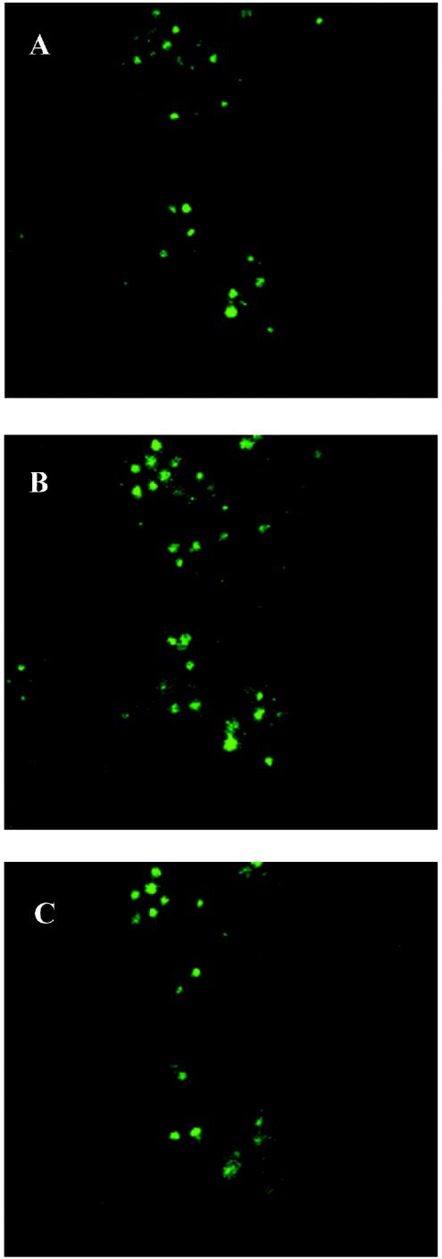
FIG. 1.
Confocal laser scanning microscopy; confocal micrographs depicting the interaction of M. hominis with T. vaginalis strain MPM2. Mycoplasmas were anti-M. hominis and FITC labeled. Optical sections were taken from the apical surface (A) of the protozoan cells, moving downward toward the basal surface (C). Section B was taken in the equatorial region of trichomonad cells and shows specific internal fluorescence.

