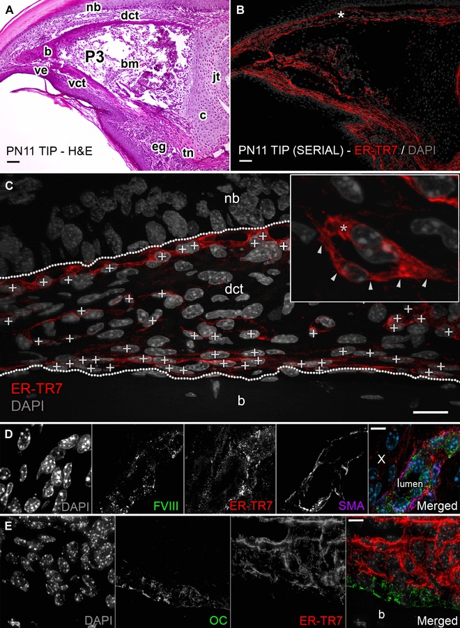
Figure 1.

ER‐TR7 outlines tissue compartments of the digit. (A) H&E section of PN11 mouse digit tip shows compartments including nail bed (nb), ventral epithelium (ve), eccrine glands (eg), and a P3 rudiment composed of both cortical bone (b) and a proximal cartilaginous (c) growth plate. P3 encloses bone marrow (bm) and ends at the P3−P2 synovial joint (jt). P3 is connected to the proximal musculature through a tendon (tn) and is surrounded by loose dorsal and ventral CT (dct and vct). (B) Adjacent section from (A) stained against ER‐TR7. (C) Representative area captured at 400× from the dct in (B) (white asterisk). The boundary landmarks of the CT (labeled nb and b) are outlined with white dotted lines. ER‐TR7+ FRCs are marked (white + signs on nuclei) and these were discriminated (C, inset) at 1000× magnification by ER‐TR7 expression in membrane extensions (white arrows) or cytosol (white asterisk) of individual cells. Scale bars (A), (B) 50 μm and (C) 25 μm. Serial sections were also co‐immunostained for (D) ER‐TR7, FVIII, and SMA (white × marks negative cells) or (E) ER‐TR7 and osteocalcin OC; scale bars (D)−(E) 10 μm
