
Figure 2.
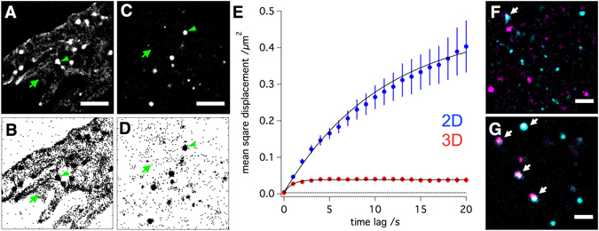
2D versus 3D cell culture conditions have a strong impact on the nanoscale distribution of integrins. (A) Normalised gaussian and (B) Scatter plot visualization of single molecule localization data obtained through a live-cell immunostaining of 2D cultured cells with a directly labeled integrin β1 antibody. Scale bar is 2 µm. (C and D) Corresponding data of a live-cell integrin β1 immunostaining of 3D cultured cells. A much higher fraction of integrins in 2D cultured cell is not part of a cluster as compared to 3D cultured cells (immobile integrin clusters = green arrowheads, individual mobile integrins = green arrows). Single molecule tracking of live cell antibody stained β1 integrins confirms this visual observation. Whereas the mean square displacement plot of β1 integrin mobility measured in 3D cultured cells shows a strong confinement (fast saturation of the curve) and low mobility (low slope, Fig. 2E, red), β1 integrins in 2D culture cells are, on average, much faster and far less confined in their mobility (Fig. 2E, blue). Fitting both plots to a model for confined diffusion reveals that β1 integrins in 2D and 3D cultured cells are confined in their mobility to an area of 1.2 ± 0,045 and 0.33 ± 0,005 µm2 with diffusion coefficients of 0,02 ± 0,0005 and 0,002 ± 0,0001 µm2/s, respectively. The dotted line represents the lower limit of the detectable square displacement (in our case 4× (28 nm)2 = 0,003136 µm2). (F) Colocalization of β1 (cyan) and β3 (magenta) integrins in 2D and (G) 3D cultured cells. Scale bar is 1 µm. Arrows indicate integrin-subunit colocalization (white) of β1 (cyan) and β3 (magenta) integrin subunits.