
Figure 7.
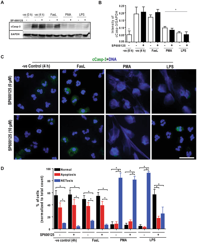
Inhibition of JNK in LPS-treated neutrophils does not lead to apoptosis, and maintains cell survival. (A–B) Immunoblot analyses of the cleave caspase-3 (cCasp-3) show that neutrophils treated with media control for 4 hours in the presence or absence of SP600125 undergo apoptosis to the extent similar to that of Fas-L treatment. Neutrophils activated by 25 nM PMA and 25 μg/ml LPS mostly show NETosis. Treating neutrophils with LPS and SP600125 shows no cCasp-3; NETosis in these cells are very low (*p < 0.05; One-way ANOVA with Tukey’s multiple comparison post test). (C) Confocal microscopy of the neutrophils stained with DAPI (blue) and cCasp-3 (green) after 4 hours. Images show some of the non-activated control neutrophils stain for cCasp-3, in the presence or absence of SP600125. Treating neutrophils with LPS, PMA and PMA with SP600125 exclusively show signs of NETosis; only traces of cCasp-3 is visible in a few of these cells. Treating neutrophils with LPS and SP600125 show no cCasp-3 or NETs (n = 3; scale bar 20 μm). See Supplementary Fig. S7 for the low magnification images. (D) The percentages of normal, apoptotic and NETotic cells were calculated based on cCasp3 staining and nuclear morphology. The quantitative analysis confirms the qualitative analysis (n = 3; *p < 0.05; One-way ANOVA with Tukey’s multiple comparison post test). Error bars in all the panels represent SEM. Collectively, inhibition of JNK activation during LPS-mediated activation of neutrophils result is cell survival.