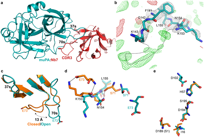
Figure 5.

The muPA:Nb7 complex crystal structure. (a) Overall structure of the serine protease domain of muPA (teal) in complex with Nb7 (red). (b) Superposition of the structure around the 140s loop in muPA:Nb7 with the 2F o–F c and F o–F c electron density maps at contour level σ = 1 (grey) and σ = ±3 (+green, −red) respectively. Black dashed line indicates a potential backbone trace of the 140s loop. (c) Structural comparison of the 37s and 70s loops in muPA between the muPA:Nb22 (orange) and muPA:Nb7 (teal) structures. (d) Superposition of the 70s/140s polar interaction network in muPA:Nb22 (orange) and muPA:Nb7 (teal) represented by sticks. (e) Structural comparison of the catalytic residues in muPA between the muPA:Nb22 structure (orange) and the muPA:Nb7 structure (teal).
