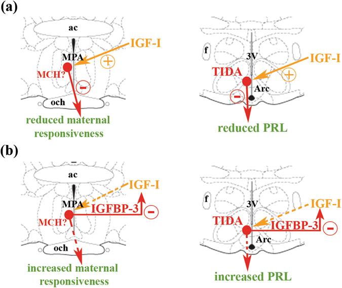
Figure 8.

A model showing how the IGF-I - IGFBP-3 system regulates maternal responsiveness and prolactin release. (a) In virgin females and pup-deprived mothers, IGF-I of hypothalamic origin or from the circulation probably induce the expression of MCH in the preoptic area and enhances the expression of the TH enzyme, furthermore stimulates its activation by phosphorylation in TIDA neurons. Consequently, these cells produce more MCH or dopamine, which inhibits maternal responsiveness or prolactin secretion from the pituitary. (b) In lactating mothers, MCH and TIDA neurons enhance the expression of IGFBP-3, which can neutralize IGF-I. The MCH or TIDA stimulating effect induced by IGF-I diminishes, which results in a decrease in MCH or dopamine production and concomitant increased maternal responsiveness or prolactin secretion due to its release from tonic inhibition. Abbreviations: ac – anterior commissure, Arc – arcuate nucleus, f – fornix, MPA – medial preoptic area, och – optic chiasm, 3 V – third ventricle.
