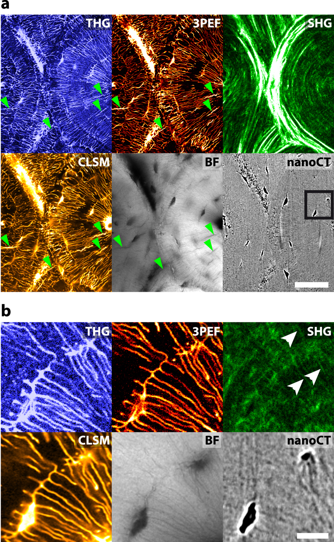
Figure 3.

Comparison of different bone imaging modalities. (a) Wide field comparison of THG (top left), 3PEF (top middle), SHG (top right), fluorescence confocal (CLSM, bottom left), brightfield transmission (BF, bottom middle) and X-ray nanoCT (bottom right) images acquired on a transverse section of a bovine femur. Some cracks are indicated by green arrowheads in (a). Additional cracks are visible on the confocal and brightfield images that were recorded after the nonlinear images. Scale bar, 50 µm. (b) Zoom in the black rectangle in (a). Collagen fibrils along some of the canaliculi are shown by white arrows on the SHG image. Scale bar, 10 µm. See also Supplementary Movie 3 and 4.
