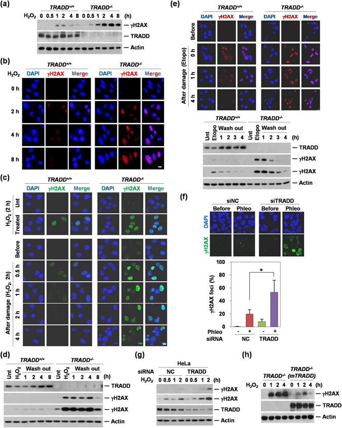
Figure 1.

Deficiency of TRADD induces impaired DNA damage response. (a) Western blotting analysis shows blotting for γH2AX, TRADD, and Actin in TRADD+/+ and TRADD−/− MEF cells treated with H2O2 in time-dependent manner (0.5 mM). (b) γH2AX foci (Red) were analyzed in TRADD+/+ and TRADD−/− MEF cells treated with H2O2 (0.5 mM) by Immunofluorescense as described in A. Scale bars, 10 μm. (c) Immunofluorescence analyses of γH2AX (Green) in H2O2 (0.5 mM) treated TRADD+/+ and TRADD−/−MEF for 2 hours (upper panels) and release from H2O2 treated TRADD+/+ and TRADD−/− MEF for 4 hours (lower panels). Cells were stained with anti-γH2AX (Green) and DAPI (Blue). Scale bars, 10 μm. (d) Western blotting analysis shows results consistent with immunofluorescence as described in (c). (e) After cells were treated with etoposide (25 μM) for 1 hour, TRADD+/+ and TRADD−/−MEF replaced with fresh media. Cells were stained with anti-γH2AX (Red) and DAPI (Blue). Western blotting analysis (lower panel) shows the consistent results with immunofluorescence. Scale bars, 10 μm. (f) Quantitative analysis of γH2AX foci was conducted in TRADD knock-downed U2OS cells. After TRADD knockdown, cells were treated with phleomycin (Phleo) and then stained with γH2AX antibody. *P < 0.05 (Student’ s t-test). (g) Transient knockdown of TRADD induces unrepaired DNA damage in HeLa cells. Western blot analysis shows γH2AX status in response to H2O2 in TRADD KD HeLa cells. Cells were transfected with siRNA TRADD or siRNA negative control (NC), respectively. After 48 hours, the cells were continuously treated with H2O2 (0.5 mM). The whole cell lysates were analysed by western blot as using indicated antibodies. (h) Reconstitution of TRADD in TRADD−/− MEFs. Western blotting analysis shows γH2AX expression in response to continuous treatment with H2O2 (0.5 mM) in different time points.
