Figure 2.

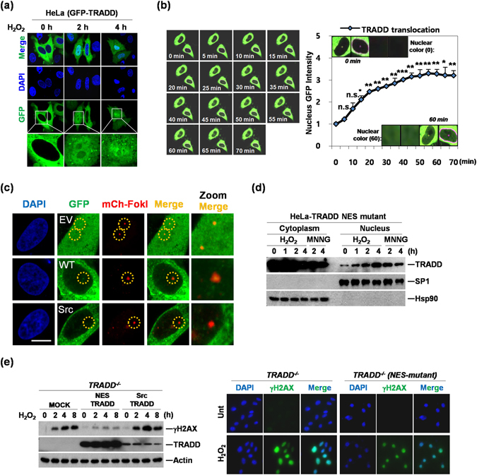
DNA damage induces nuclear translocation of TRADD. (a) HeLa cells were transiently transfected with GFP-TRADD and treated with H2O2 (0.5 mM) for indicated time points. Cells were analyzed by confocal fluorescence microscopy. (b) HeLa cells were transiently transfected with GFP-TRADD and treated with H2O2 (0.5 mM). After treatment, live cell Images were analyzed by confocal fluorescence microscopy for 70 minutes (left panel). Quantitative analysis of nuclear translocation of TRADD was measured by GFP intensity in the nucleus (right panel). *P < 0.05; **P < 0.01; ***P < 0.001; n.s., not significant (Student’s t-test). (c) Colocalization of GFP-TRADD and mCherry-FokI at single DNA double-strand break site. GFP empty vector (EV), GFP-TRADD wild type (WT), or GFP-TRADD Src mutant (SRC) was cotranfected with mCherry-FokI (mCh-FokI) nuclease into U2OS 2-6-3 cell lines. After 48 hr, cells were fixed and stained with DAPI for nuclear staining. Images were analyzed confocal microscope (Nikon A1). Scale bar, 10 μm. (d) HeLa cell were transiently transfected with NES mutant TRADD and treated with H2O2 (0.5 mM) or MNNG (0.25 mM) for indicated times. Cells were fractionated into cytoplasmic and nuclear fractions using an NE-PER fractionation kit. Anti-Hsp90 or anti-Sp1 used as a control for normalization of cytoplasm and nuclear lysates, respectively. (e) Western blotting analysis was conducted with lysates from TRADD−/− (MOCK), NES-mutant TRADD (NES-TRADD) and Src-myristoylation-TRADD (Src-TRADD) in TRADD−/− MEFs treated with H2O2 (0.5 mM) for indicated time periods (left panel). Expression of γH2AX was analyzed in TRADD−/− and TRADD−/− (NES-mutant TRADD) MEFs treated with H2O2 (0.5 mM) for 1 hour using immunofluorescence (right panels).
