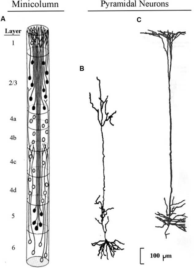
FIGURE 1.

Pyramidal neurons with long apical dendrites from layers 5 and 6. (A) Minicolumn diagram adapted from Peters and Sethares (1991) of a slice of monkey visual cortex, which shows locations of somas and apical dendrites (basal dendrites and axons are omitted). Somas showing no attached fibers are stellate neurons, which contain many short dendrites that radiate from the soma in all directions (not shown here). The vertical length of the minicolumn indicates the thickness of the cortical area in which the two pyramids were observed. (B) Camera lucida drawing of a layer 6 pyramidal neuron of the monkey from Figure 4 of Wiser and Callaway (1996); (C) Camera lucida drawing of a layer 5 pyramidal neuron of the monkey from Figure 24 of Valverde (1986).
