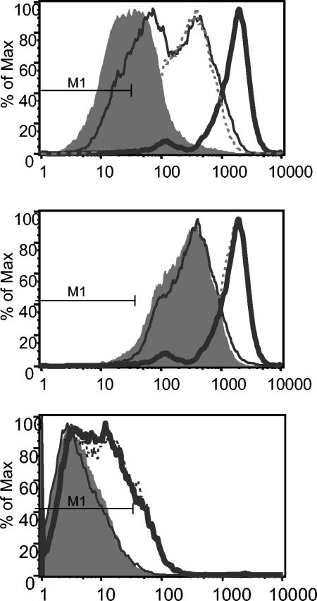
FIG. 7.
Adhesion molecule expression by HUVEC after H. pylori-induced TEM. PBMC were allowed to migrate toward H. pylori strain Hel 312, urease, or the 26-kDa protein in the Transwell system, and investigation of ICAM-1 (top and middle panels) and VCAM-1 (bottom panel) expression on HUVEC after TEM was performed by using flow cytometry. The top panel shows ICAM-1 expression with or without PBMC and H. pylori added. The filled histogram shows ICAM-1 expression on untreated HUVEC, the thin solid line shows ICAM-1 expression after addition of H. pylori alone, the dashed line shows ICAM-1 expression after addition of PBMC alone, and the bold solid line shows ICAM-1 after the addition of both H. pylori and PBMC for 16 h, allowing TEM. The middle and bottom panels show ICAM-1 and VCAM-1 expression on HUVEC after transmigration of PBMC toward medium (filled histogram), Hel 312 (bold solid line), urease (dashed line), or 26-kDa protein (thin solid line). M1 indicates the fluorescence of the isotype control in all histograms.

