
Figure 7. OSBP1 mediates SL flow‐induced PtdIns(4)P consumption.
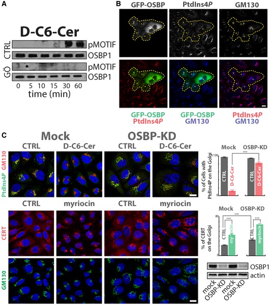
- Cells expressing OSBP1‐GFP were treated for the indicated times with D‐C6‐Cer (10 μM) and lysed, and protein lysates were subjected to immunoprecipitation by the use of anti‐GFP antibody, SDS–PAGE and Western blotting. PKD‐dependent phosphorylation of OSBP was revealed by pMOTIF antibody. The PKD inhibitor Gö 6976 (10 μM, pre‐treatment for 1 h) was used as a specificity control.
- Cells transiently expressing OSBP1‐GFP were fixed and permeabilized as in Fig 3 and stained with anti‐GM130 (blue) and anti‐PtdIns(4)P (red). Asterisks and dashed line indicate OSBP1‐GFP‐transfected cells.
- Cells KD for OSBP1 were treated with D‐C6‐Cer (10 μM) for 1 h, fixed, permeabilized and stained as in Fig 3A (upper left panels). The percentage of cells showing PtdIns(4)P at the Golgi in the different conditions tested is reported (upper right panel). Cells KD for OSBP1 were treated with myriocin (2.5 μM for 24 h), fixed, permeabilized and stained for CERT (red) and GM130 (green). CERT recruitment to the Golgi region was evaluated by immunofluorescence and quantitated as in Fig 1C (middle right panel). OSBP1 KD efficiency was evaluated by Western blotting (lower right panel). ***P < 0.001; according to two‐tailed Student's t‐test.