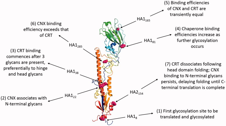
Figure 4.
The role of N-linked glycosylation in the folding of INFV HA. The figure shows HA of the INFV A/Aichi/68-derived X31 strain (H3N2) with data derived from PDB ID: 1HGF (Sauter et al., 1992b). The polypeptide chain is colored in an N- to C-terminal blue-to-red gradient. The asparagine residues of the seven N-linked glycosylation sites are highlighted in magenta spheres and numbered according to position within mature HA1 or HA2. Labels indicate how the binding of CNX and CRT varies during cotranslational glycosylation.

