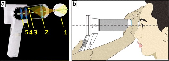
Fig. 2.

(a) Optical configuration of the prototype camera (distances between components are approximated). The components are specified as follows: (1) human eye, (2) front objective lens, (3) condensing lens, (4) visible light emitting diode (LED) unit and xenon flash tube, (5) macro lens. This configuration allows a reflection-free, 60° field–of-view of the fundus in an external housing that can be attached to an LCD monitor with a CMOS sensor that maintains hand-held, point-and-shoot operation. (b) Operation of the non-mydriatic portable fundus camera while screening retinal diseases
