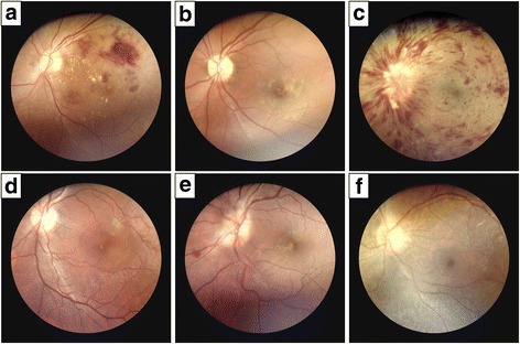
Fig. 5.

Fundus photographs obtained with the DEC200 portable fundus camera. The unilateral fundus photographs of the study participants show a variety of retinal pathology, including (a) diabetic retinopathy with exudate and hemorrhage, (b) geographic atrophy with drusen, (c) central retinal vein occlusion (CRVO), (d) central serous chorioretinopathy, (e) papillitis, and (f) retinitis pigmentosa (RP)
