
Fig. 3.
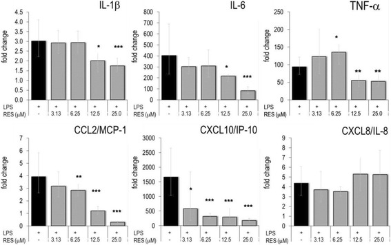
Effect of RES on gene expression in activated THP-1. PMA-treated THP-1 cells were cultured in the presence of indicated concentrations of RES and activated with LPS for 4 h. RT-PCR was performed and the gene expression levels were indicated as mean fold changes (± errors) (versus unstimulated cell) (see also reference [11]). * p < 0.05, ** p < 0.01, *** p < 0.001 (versus LPS-stimulated cells)