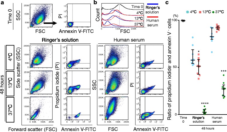
Fig. 4.

Apoptosis of synovial MSCs 48 h after preservation. a Representative profiles of synovial MSCs by forward scatter (FSC) and side scatter (SSC), stained with fluorescein isothiocinate (FITC)-annexin V and propidium iodide (PI). Gates were placed around major cell populations. b Representative forward scatter histograms. c Apoptotic to normal cell ratios. Cells negative for annexin V and propidium iodide were considered nonapoptotic cells. Median values and interquartile ranges are shown (n = 3). *p < .05, ***p < .001, ****p < .0001, compared with the value at Time 0 by Kruskal-wallis test followed by Dunn’s multiple comparisons
