Fig. 7.

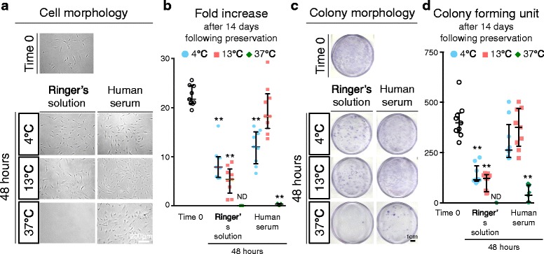
Fold increase and colony formation unit of synovial MSCs before and 48 h after preservation. Passage 2 synovial MSCs before and 48 h after preservation were replated and cultured for 14 days. a Representative cell morphologies of synovial MSCs. b Fold increases after 14 days in culture before and 48 h after preservation. Median values and interquartile ranges are shown (n = 3). c Representative dishes stained with crystal violet. d Colony forming units after 14 days in culture before and 48 h after preservation. Median values and interquartile ranges are shown (n = 3). **p < .01, compared with the value at Time 0 by Friedman test followed by Steel’s multiple comparisons. ND not detected
