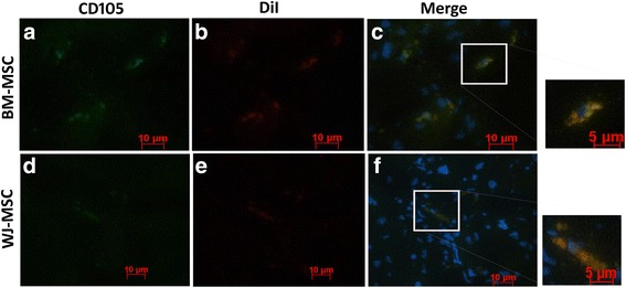
Fig. 5.

Immunofluorescence analysis for a–c BM-MSCs and d–f WJ-MSCs engraftment in the liver tissues of CCl4-treated rats after 30 days of cell injection. Representative fluorescence images show colocalization of human CD105 expression (a, d) and DiI-positive cells (b, e) in liver tissue sections. The photomicrographs were captured using 40× and 60× (insets) objectives. BM-MSCs; bone marrow-derived mesenchymal stromal cells, WJ-MSCs; Wharton’s jelly-derived mesenchymal stromal cells
