
Fig. 2. Iron oxide nanoparticle characterization.
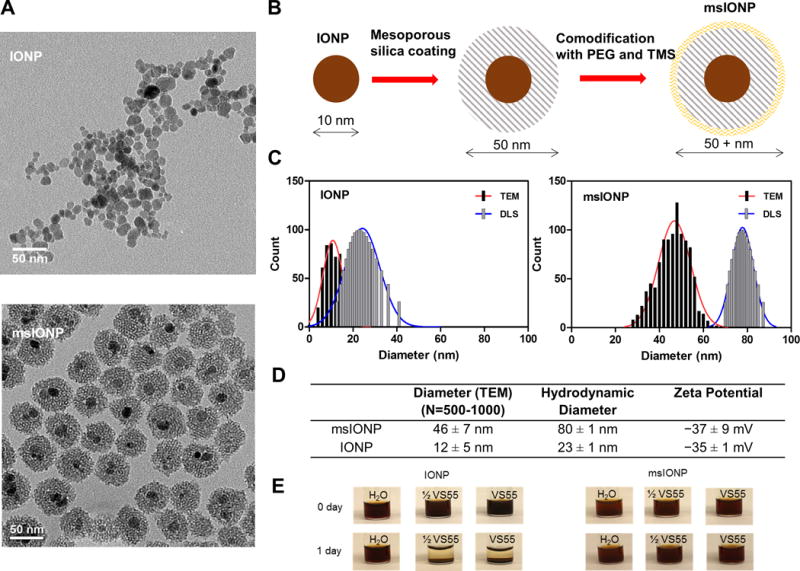
(A) Representative TEM images of IONPs and msIONPs. (B) Schematic detailing the synthesis of msIONPs. The IONPs were coated with mesoporous silica shell followed by co-modification of PEG and TMS on the surface of msIONPs. (C) Size distribution of IONPs and msIONPs quantified by dynamic light scattering (DLS) and by analyzing 500 − 1000 NPs from TEM images. (D) Table of parameters of IONPs and msIONPs. (E) Photographs depicting stability of msIONPs and IONPs in VS55 at room temperature over time.