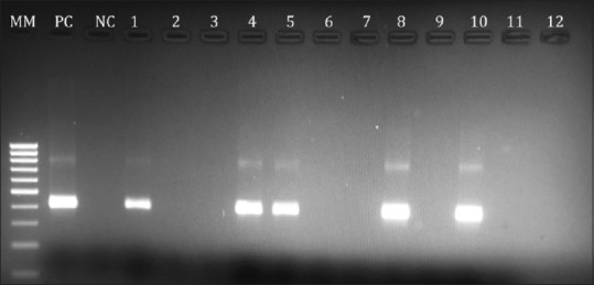
Figure 1.

Agarose gel electrophoresis photo showing the HDV PCR products; 1st lane (MM) 100bp ladder marker; 2nd lane positive control (PC) for HDV; 3rd lane negative control (NC); lanes no. 1,4,5,8 and 10 are positive HDV PCR samples with product size 404 bp and lanes 2,3,6,7,9,11 and 12 are negative HDV PCR samples
