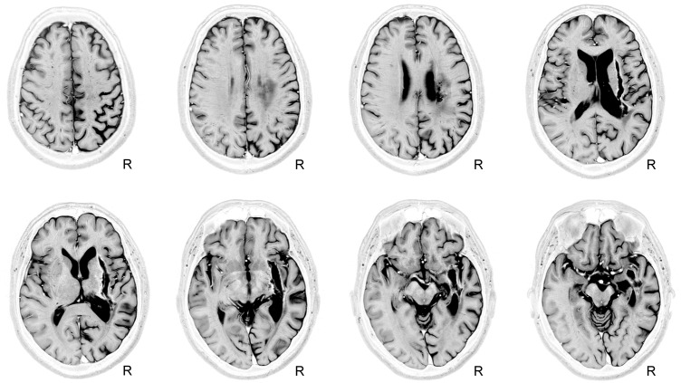
FIGURE 2.
Depiction of an old right subcortical hemorrhage on a Short-TI Inversion Recovery (STIR) MRI sequence. Axial views of MRI-STIR images are shown in native space. The MRI shows an extensive lesion with a semilunar configuration involving the right striatum-capsular region extending into the surrounding white matter. Mild post-stroke right temporal-parietal cortical atrophy is evident. See text for further details. The neurological convention is used. R, right.

