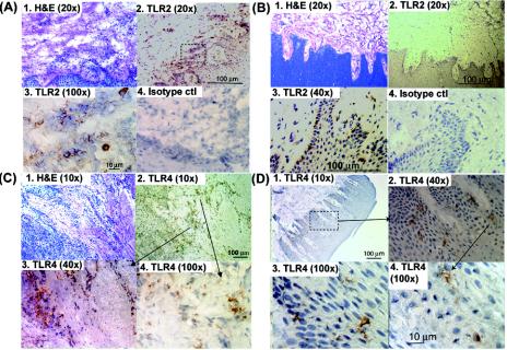
FIG. 1.
Oral mucosa infiltrated with TLR2+ and TLR4+ cells. Representative serial sections of interproximal gingival tissue from patients with CP (A and C) persons in relative health (B and D) were single-immunoenzyme stained as described in Materials and Methods. Images of TLR2+ cells (A and B) and TLR4+ cells (C and D) were obtained with a 20× objective (panels A1, A2, B1, and B2), a 40× objective (panels B3, B4, C3, and D2), a 10× objective (panels C1, C2, and D1), 100× objective (panels A3, C4, D3, and D4) and captured using image-enhanced light microscopy. The specificity of the antibodies was confirmed by negative staining with the respective isotype controls (e.g., panels A4 and B4). The sections were counterstained with hematoxylin.

