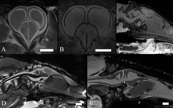
Fig 1. MR images of crocodilian brains.
T2w coronal image of an embryo—scan time 3 h 21m (A), T1w coronal image of an early juvenile—scan time 9 h 6 m (B), T1w sagittal image of a late juvenile—scan time 2 h 36 m (C), T2w sagittal image of an adult—scan time 7 m (D), T1w sagittal image of an adult—scan time 40 m (E). Scale bar– 5 mm (A, B, C) and 10 mm (D, E). Double-headed arrows in (E) show the typical area of the interstitium.

