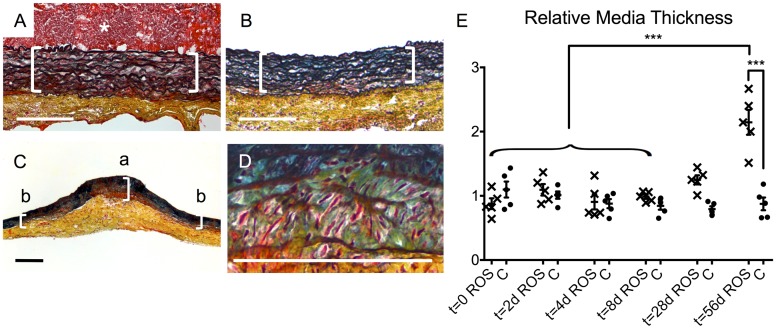
Fig 7. Histomorphological changes of the media.
(A) A representative cross-section image of the ROS group displayed intact organization of closely spaced laminae of the media (brackets) at t = 0. The same appearance was found in the ROS specimens after thrombus-dissolution at t = 28 d (B) and in the controls at all times. (C) In contrast, ROS specimen showed at t = 56 d a distinctive, media-pronounced wall thickening at the treated focus (bracket “a”) compared to non-treated areas of the same specimen (brackets “b”). (D) A detailed analysis of this focus revealed a loosening of the laminae and enlarged amounts of green-bluish intercellular substance, consisting of high proportions of proteoglycans. Movat´s pentachrome staining: collagen (yellow), fibrin (red), proteoglycans (blue-green), elastin (black), nuclei (blue to black), cytoplasm (pink); asterisk = thrombus; scale bars = 200 μm. (E) Quantitative analysis of the media thickening at the irradiated focus. ROS = ROS group; C = control group; *** p < 0.001.

