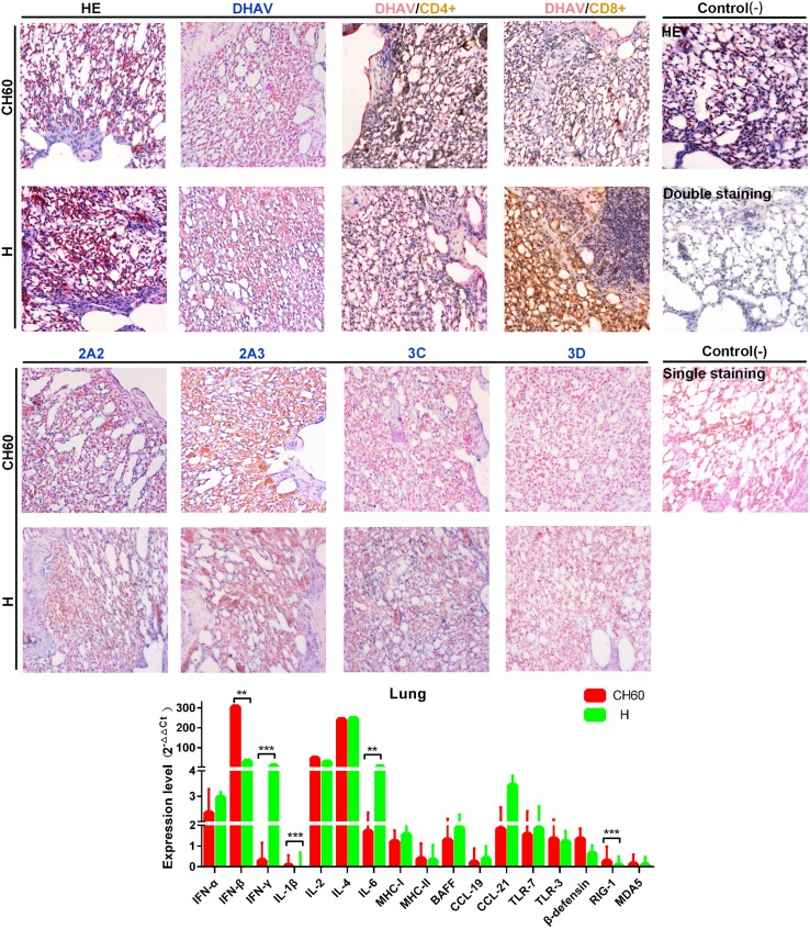
Fig 10. Comparative study of virus-host interactions mediated by attenuated and virulent strains in the lung.
HE, single staining of the viral capsid, 2A2, 2A3, 3C and 3D and double staining of the viral capsid and CD4+ or CD8+ positive cells were performed to compare the pathological changes, viral protein expression levels and the Th or Tc cell responses caused by the diversity of virulence. The positive staining of the viral capsid, 2A2, 2A3, 3C and 3D was visualized as bluish violet, while the double staining of the viral capsid and CD4+ or CD8+ was identified as red or brown, respectively. The negative control (-) refers to standard HE staining and single and double IHC staining without primary antibody. The relative expression levels of immune-related genes were calculated using the 2-ΔΔCt method. The error bars represent the standard errors of the mean. Statistically significant differences induced by attenuated and virulent strains between the mean gene expression levels were determined by one-way ANOVA. *P < 0.05, **P < 0.01 or ***P < 0.001 indicate the level of statistical significance of differences between the different groups.

