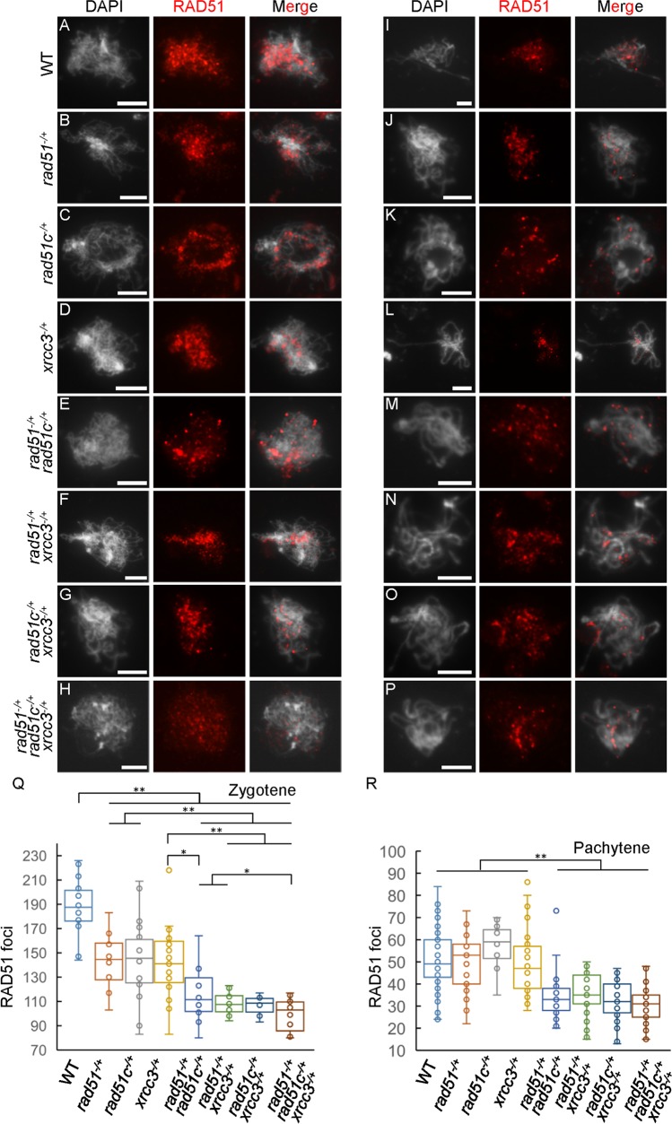
Fig 6. Immunostaining of AtRAD51 signals in single, double and triple heterozygous mutant chromosomes at zygotene and pachytene.
(A-H) The locations and (Q) numbers of AtRAD51 foci on zygotene chromosomes from atrad51-/+, atrad51c-/+, atxrcc3-/+, atrad51-/+ atrad51c-/+, atrad51-/+ atxrcc3-/+, atrad51c-/+ atxrcc3-/+ and atrad51-/+ atrad51c-/+ atxrcc3-/+ heterozygous mutant meiocytes showed reductions compared to wild type (WT). (I-L) The location and (R) number of AtRAD51 foci on pachytene chromosomes in the three single heterozygotes showed no obvious differences compared with WT. (M-P) The location and (R) number of AtRAD51 foci on pachytene chromosomes in the double and triple heterozygous mutants were significantly reduced relative to WT. Left panels show the chromosome morphology following staining with 6-diamidino-2-phenylindole (DAPI), middle panels show AtRAD51 foci (red dots), and right panels merge the DAPI-stained images with the AtRAD51 foci images. Scale bar: 5 μm. ** p<0.01 (two-tailed Student’s t-test).

