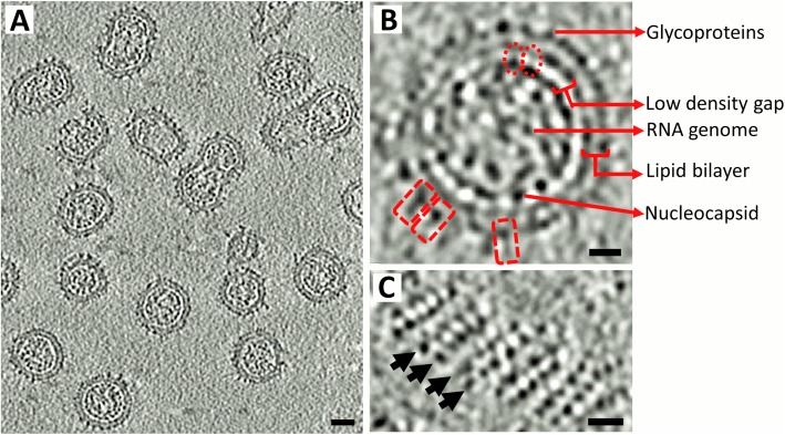
Fig 1. Rubella virion morphology.
(A) Section from a rubella virus tomogram showing the different morphologies of rubella virions. Scale bar corresponds to a length of 200 Å. (B) Cross-section of a rubella virion. The dashed rectangles in red show individual rubella glycoprotein spikes. The dashed ovals in red indicate thin densities that connect the inner nucleocapsid shell to the outer glycoprotein plus membrane shell. (C) Surface of a rubella virion showing the glycoprotein rows. Black arrows mark the direction of the rows. Scale bar in panels B and C correspond to a length of 100 Å. Black is high density in all panels.

