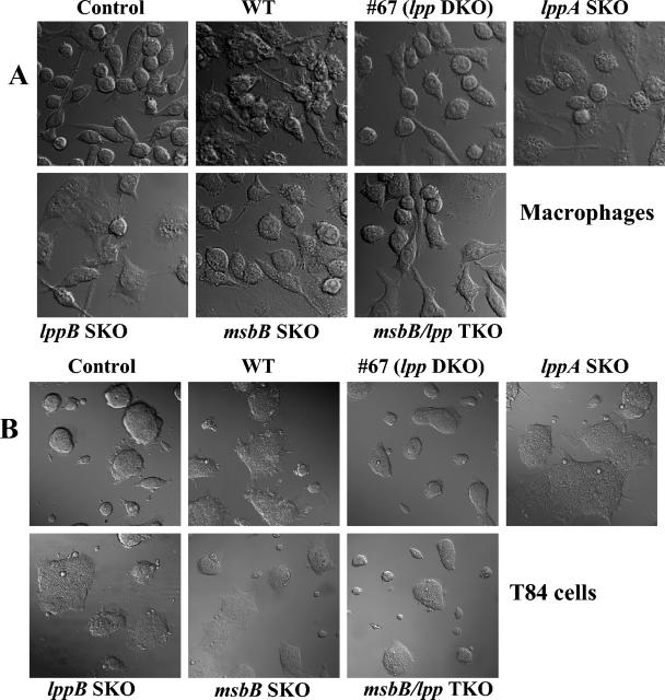
FIG. 1.
Cell death induced by WT S. enterica serovar Typhimurium and S. enterica serovar Typhimurium mutants in RAW264.7 (A) and T84 (B) cells. Cells were infected with various S. enterica serovar Typhimurium mutants for 1 h at an MOI of 10:1. After incubation, the cells were washed with PBS and incubated in a gentamicin (100 μg/ml)-containing medium for 1 h. After washing, the cells were incubated in an antibiotic-free medium for 24 h. The cells were gently washed in PBS and examined with a laser scanning confocal microscope. Note the normal cell morphology of noninfected cells (control) and of cells infected with the lpp DKO mutant (#67) (75% viability) and the lpp-msbB TKO mutant (mostly normal) compared to the cell morphology of dead cells after infection with the WT and the lppA and lppB SKO mutants. The msbB SKO mutant-infected host cells showed around 30% destruction. At least 20 microscopic fields were examined.

