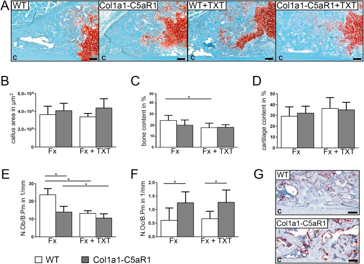
Fig 3. Fracture healing in WT and Col1a1-C5aR1 mice 14 d after surgery.
(A) Representative histological images of wildtype (WT) and Col1a1-C5aR1 mice with isolated fracture and fracture with additional thoracic trauma (TXT). (B) Callus area, (C) amount of osseous tissue and (D) amount of cartilage. (E) Number of osteoblasts per bone perimeter (N.Ob/B.Pm) and (F) number of osteoclasts per bone perimeter (N.Oc/B.Pm) of WT and Col1a1-C5aR1 mice. (G) Representative tartrate-resistant acid phosphatase (TRAP) staining of fractured calli of WT and Col1a1-C5aR1 mice with isolated fracture. Fx: mice with isolated fracture, Fx+TXT: mice with combined fracture and thoracic trauma. C: cortex, scale bar 100 μm, *p < 0.05, n = 6 per group.

