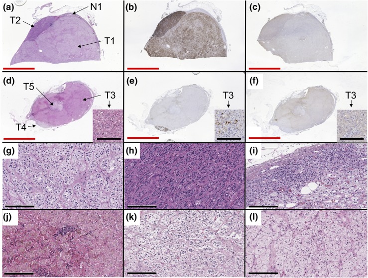
Figure 1.
Images of histology and IHC. (a and d) Hematoxylin and eosin, (b and e) CYP11B2 IHC, (c and f) CYP17A IHC, and high magnitude images (200×) of (g) T1, (h) T2, (i) N1, (j) T3, (k) T4, and (l) T5. One block (a–c) showed two collision adenomas consisting of ZF-like cells (g) and ZG-like cells (h) and adjacent normal adrenal tissue with paradoxical hyperplasia (i). The other block (d–f) showed three collision adenomas consisting of ZR-like cells (j) and ZF-like cells (k and l). Some cells in T3 were positive for CYP11B2 and CYP17A IHC, as shown in small pictures in (d–f) (note the same structure of interstitial tissues arising from left bottom to right middle). Red and black bars indicate 10 mm and 200 µm, respectively.

