
Fig. 1. S. elongatus successfully cocultured with rat cardiomyocytes.
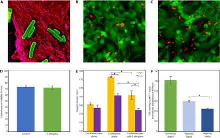
(A) False-colored scanning electron micrograph of multiple S. elongatus cyanobacteria (green) with a single rat cardiomyocyte (red). (B and C) Representative images of a live/dead assay of cardiomyocytes alone (B) and cardiomyocytes cocultured with S. elongatus (C). The cytoplasm of live cells is stained green, and the nuclei of dead cells are stained red; the autofluorescent rod-shaped S. elongatus are distinct from the circular dead nuclei. (D) Results of the live/dead assay demonstrating that the presence of S. elongatus does not affect cardiomyocyte survival. (E) In vitro oxygen measurements of cardiomyocytes alone, S. elongatus alone, and cardiomyocytes cocultured with S. elongatus under light and dark conditions in normoxia. In cultures containing S. elongatus alone and S. elongatus with cardiomyocytes, oxygen tension was significantly higher in the presence of light (P < 0.01 and P = 0.03, respectively), suggesting active photosynthesis. Cardiomyocytes cocultured with S. elongatus in the light demonstrated a positive trend toward increased oxygen when compared to cardiomyocytes alone (P = 0.1). When compared to cultures with S. elongatus alone, cultures with cardiomyocytes and S. elongatus demonstrated significantly reduced oxygen in light and dark (P = 0.01 and P < 0.01, respectively), suggesting oxygen consumption by cardiomyocytes. (F) Cellular viability was assessed using a WST-1 cell proliferation assay. Cardiomyocytes cultured with S. elongatus demonstrated significantly enhanced cellular metabolism in the presence of light as compared to dark (P < 0.01). All data are reported as means ± SEM. *P < 0.05 using two-tailed unpaired Student’s t test.