
Fig. 2. Reduced deformability of primed neutrophils in the OS.
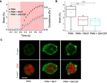
(A) Representative strain and compliance time course (mean ± SEM) from n ≥ 4 experiments for resting (n = 56), fMLP-treated (n = 24), and GM-CSF–treated (n =39) neutrophils. Reduction in strain and compliance is seen in GM-CSF– and fMLP-treated cells. (B) fMLP- and GM-CSF–treated neutrophils are significantly stiffer than resting cells [****P < 0.0001 and ***P < 0.001 (significant difference)]. (C) Confocal images of Alexa Fluor 488 Phalloidin–stained cells showing F-actin distribution in fixed resting and chemically activated neutrophils. Scale bar, 5 μm.