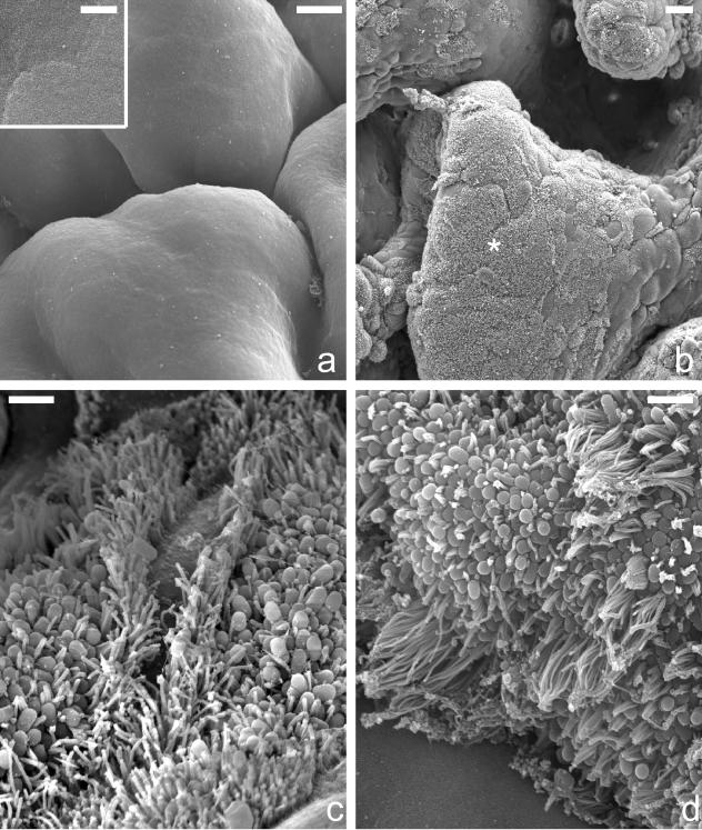
FIG. 1.
Scanning electron micrographs showing uninfected duodenal mucosa (a) and mucosa infected for 8 h with wild-type E2348/69 (b and c) and E2348/69 bfpA mutant strain UMD901 (d). Control tissue displayed a uniformly smooth mucosal surface with the outline of individual enterocytes visible, although even at high power, brush border microvilli were not resolved (a, inset); uninfected mucosa lacked any adherent bacteria (a). Following an 8-h infection with E2348/69, a large percentage of the mucosal surface was colonized by adherent bacteria (b, asterisk). Adherent bacteria had produced A/E lesions, and there was gross microvillous elongation, particularly around the periphery of bacterial microcolonies (c). Similar features were seen with the bfpA mutant strain (d). Size bars: a, 5 μm (inset, 0.5 μm); b, 10 μm; c and d, 1 μm.

