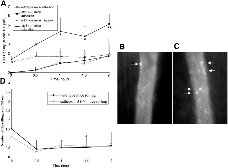
Figure 7. Leukocyte behavior in the in vivo microvasculature of ctsB-deficient mice.
(A) The number of carboxyfluorescein succinimidyl ester-labeled leukocytes (number of cells per millimeter of vessel length) adhered to the endothelium of postcapillary venules and migrating into the tissues in the cremaster-muscle microcirculation of WT mice and ctsB−/− mice after superfusion, including 10 nM PAF. There is no significant difference in flow velocity between the 2 groups. *P, #P, and **P < 0.05 vs. unsheared group at each time point. n = 4 animals in each group. (B and C) Micrographs of labeled leukocytes in inflamed postcapillary venules of cremaster muscle in a WT mouse (B) and a ctsB−/− mouse (C) under normal physiologic levels of flow rates. The muscle is superfused, including 10 nM PAF for 1 h. In the WT mouse, a few adherent leukocytes are found (arrows). In the ctsB−/− mouse, more fluorescent-labeled leukocytes adhering to vascular wall are consistently present (arrows). (D) Rolling leukocyte behavior in vivo in the inflamed skeletal muscle microcirculation of WT mice and ctsB−/− mice. The number of leukocytes rolling across a predetermined vessel cross-section was counted during a 30-s period.

