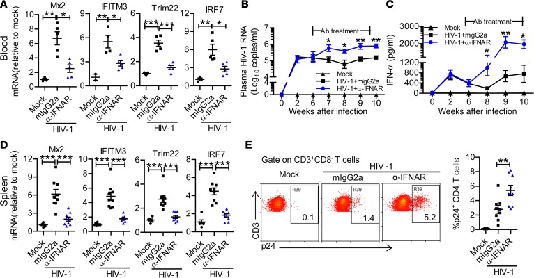
Figure 3. IFNAR1 blockade during persistent HIV-1 infection enhances viral replication in humanized mice.
Humanized mice infected with HIV-1 were treated from 6 to 10 weeks postinfection (wpi) with a mAb against IFN-α/β receptor 1 (IFNAR1) or isotype control (mouse IgG2a) twice per week. (A) Relative mRNA levels of human interferon-stimulated genes including Mx2, IFITM3, TRIM22, and IRF7 in peripheral blood mononuclear cells at 9 wpi. (B) Plasma HIV-1 RNA levels at indicated time points after HIV-1 infection. (C) Human IFN-α levels in the plasma after HIV-1 infection. Shown (A–C) are representative data (mock, n = 3; HIV-1 + mIgG2a, n = 5; HIV-1 + anti-IFNAR1, n = 5 ) of 3 independent experiments (mock, n = 7; HIV-1 + mIgG2a, n = 11; HIV-1 + anti-IFNAR1, n = 12 in total) with mean values ± SEM. (D) Relative mRNA levels of human Mx2, IFITM3, TRIM22, and IRF7 in splenocytes at 10 wpi. (E) Representative FACS plots and summarized data show the percentages of HIV-1 p24–positive CD4+ T cells (CD3+CD8–) in the spleen at 10 wpi. Shown (D and E) are combined data of 2 independent experiments (mock, n = 6; HIV-1 + mIgG2a, n = 9; HIV-1 + anti-IFNAR1, n = 9) with mean values ± SEM. *P < 0.05, **P < 0.01, ***P < 0.001. One-way ANOVA and Bonferroni’s post hoc test were performed to compare between groups at singular time points (B and C) or between groups (A, D, and E).

