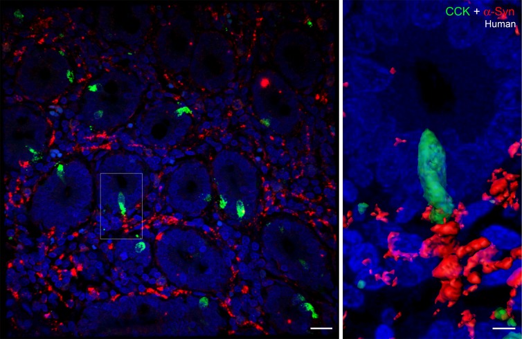
Figure 3. α-Synuclein expression in CCK cells and enteric nerves of human duodenum.
Paraffin-embedded section (5-μm thickness) of human duodenum showing a cross section of villi. α-Synuclein (red) staining is visible in enteric nerves located between the villi. One of the cholecystokinin (CCK) cells (boxed) is shown at higher magnification on the right. α-Synuclein is present inside the CCK cell, and this cell is present in close proximity to an α-synuclein–containing enteric nerve. Scale bar: 20 μm (left); 5 μm (right).

