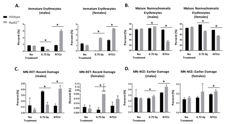
Figure 2. In vivo micronucleus assay detects genome instability in Rad52−/− mice.

Cells which are genomically unstable or mice that have been treated with a genotoxin have a higher frequency of micronucleus formation. Mouse blood samples are collected into liquid heparin solution and fixed in cold methanol. Samples are prepared and incubated in buffer containing FITC-conjugated CD71 antibody and RNase. Samples are washed and resuspended in buffer plus PI and analyzed by collecting 200,000 events by flow cytometry. Micronuclei are PI-positive, and they can be differentially identified in NCEs or RETs by co-staining with CD71. Mice are either not treated, treated with 0.75 Gray irradiation, or treated with 38 weeks of NTCU painting. A.-B. First, percentages of immature to mature erythrocytes were analyzed. C.-D. Then, DNA damage was measured as indicated by incidence of micronuclei. Micronucleated RETs are indicative of recent damage, whereas micronucleated NCEs are indicative of damage caused > 72 h earlier. P-value and significance calculated to only compare wild type v. knockout in each individual treatment group and quadrant. Multiple testing adjustments were performed so that the threshold would be less than the Bonferroni correction using p < 0.05 as threshold. *p < 0.0167 was considered to be statistically significant. Wild type mice (n = 13); Rad52−/− mice (n = 8).
