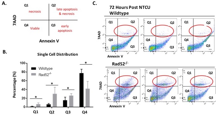
Figure 3. Changes in plasma membrane upon NTCU treatment detected by Annexin V assay.

Detection of apoptosis and necrosis by concurrent staining with annexin V-APC and 7-AAD. A. Live cells (Viable) are both annexin V and 7AAD negative. At early stage of apoptosis (early apoptosis) the cells bind annexin V while still excluding PI. At late stage of apoptosis and early necrosis they bind annexin V and stain brightly with 7AAD. In exclusively necrotic cells, they stain only 7AAD. B.-C. Wild type mouse lung cells (C. top panel) and Rad52−/− mouse lung cells (C. bottom panel) were treated with NTCU for 72 h, as described in Materials and Methods. Cells were digested with MACS lung digestion kit and subsequently stained with annexin V - APC conjugate and 7AAD and their fluorescence was measured by flow cytometry. P-value and significance calculated to compare wild type v. knockout in each individual quadrant. Multiple testing adjustments were performed so that the threshold would be less than the Bonferroni correction using p < 0.05 as threshold. *p < 0.0125 was considered to be statistically significant. Wild-type (n = 6); Rad52−/− mice (n = 6).
