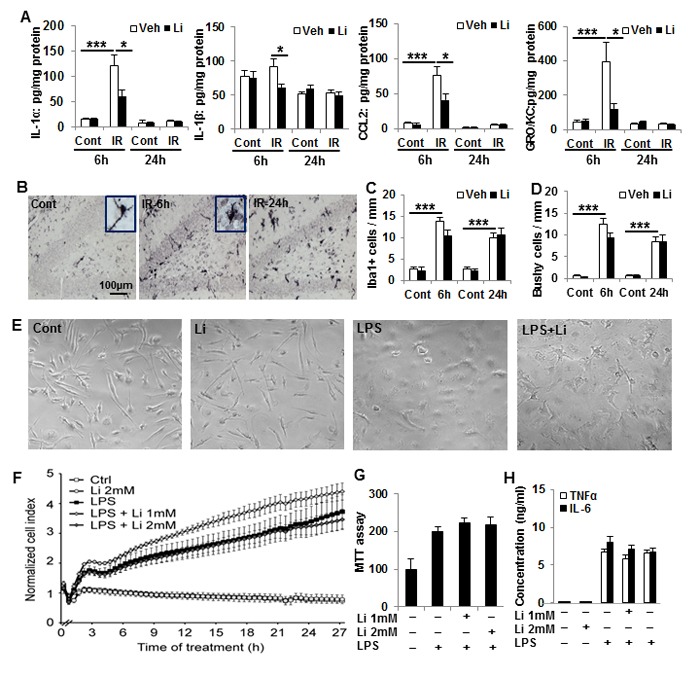
Figure 5. Lithium prevents irradiation-induced early inflammation in the hippocampus, but not effect on microglia activation in vivo and in vitro.

A. IL-1α, MCP-1, and GRO/KC increased dramatically at 6 h after irradiation compared to the non-irradiated controls. Lithium treatment prevented increases in IL-1α, MCP-1, and GRO/KC (D). The level of IL-1β was lower in the irradiated pups with lithium treatment at 6 h, but not at 24 h (n = 5 for 6 h control, n = 7 for 6 h irradiation, n = 4 for 24 h control, and n = 5 for 24 h IR). B. Representative Iba1 immunostaining of normal control and 6h, 24h after irradiation in the DG of hippocampus. C. Bar graph shows Iba1 positive cells increased significantly after irradiation compared to no-irradiated controls. D. Quantification of bushy cells based on the Iba1 staining and morphology. Most of the Iba1 positive cells were bushy morphology after irradiation. E. Representative pictures of microglia morphology in vitro under the four different conditions. F. xCELLigence analysis of microglia cells treated with LPS and/or lithium. G. Viability (MTT) analysis of microglia treated with LPS and lithium. The number and/or size of microglia increased after LPS treatment, and lithium had no effect on this. H. A bar graph of TNFα and IL-6 concentrations in the microglia culture medium. LPS stimulates TNFα and IL-6 secretion, and lithium had no apparent effect on this. *p < 0.05, ***p < 0.001.
