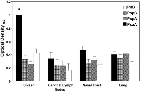
FIG. 3.
PsaA-, PspA-, PspC-, and PdB-specific proliferative responses by antigen-stimulated CD4+ T cells following pneumococcal carriage. BALB/c mice were intranasally challenged with ≈7.5 × 10⁶ CFU of Streptococcus pneumoniae EF3030 in 15 μl of Ringer's solution. Nasal tract-, lung-, cervical lymph node-, and spleen-derived CD4+ T cells were purified and antigen stimulated with 5 μg of PsaA, PspA, PspC, and PdB per ml for 3 days in complete medium. Proliferation was measured by detecting the level of bromodeoxyuridine incorporation by ELISA. The data presented are the mean ± standard error of the mean optical densities of quadruplicate cultures from infected mice. Naïve mice displayed responses that were below the detection limit of the ELISA and are not shown. Asterisks indicate significant differences (P < 0.05) between the maximum and next highest antigen-specific CD4+ T-cell proliferation response from infected mice.

