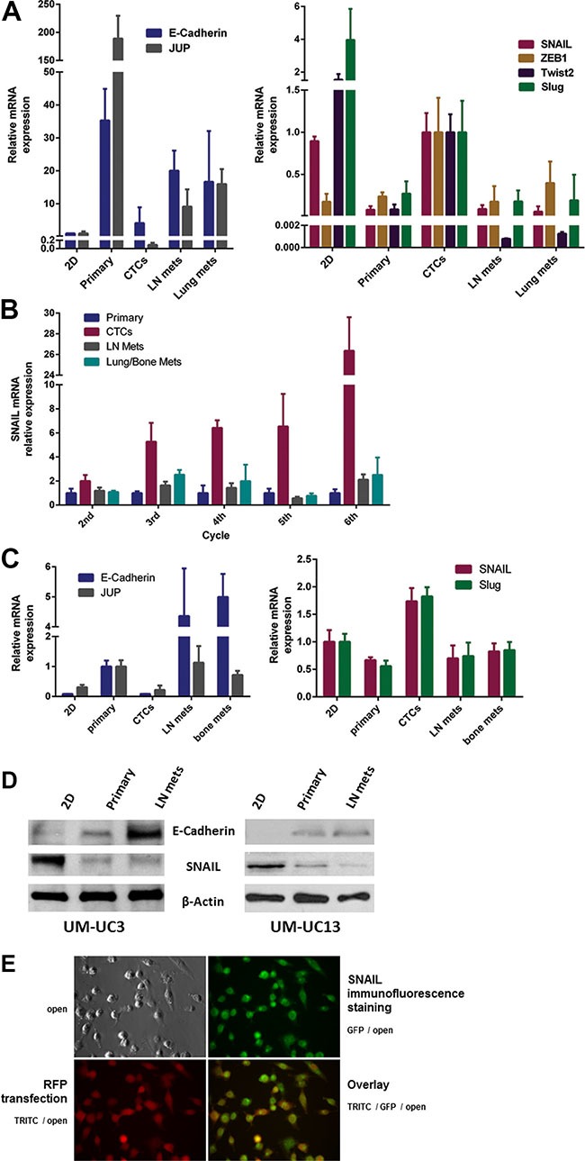
Figure 6. Expression of “epithelial” and “mesenchymal” markers in vitro and in vivo.

(A) Comparison of “epithelial” markers (left panel) and “mesenchymal” markers (right panel) for UM-UC3 in vitro (2D culture) and in vivo after the 3rd generation of recycling. Gene expression by the primary tumor, CTCs, lymph node (LN) metastases, and distant metastases were characterized by qRT-PCR. (B) Relative mRNA expression of SNAIL in UM-UC3 following successive orthotopic tumor recycling. CTCs demonstrate increasing SNAIL expression with successive generations. (C) Comparison of “epithelial” markers (left panel) and “mesenchymal” markers (right panel) for UM-UC13 in vitro (2D culture) and in vivo after the 3rd generation of recycling using qRT-PCR. (D) Immunoblots for SNAIL and E-Cadherin of 2D culture cells, primary tumors, and metastases. (E) Cell suspension staining (GFP) for SNAIL in CTCs originating from mice with UM-UC3 orthotopic tumors which are luc-RFP labelled.
