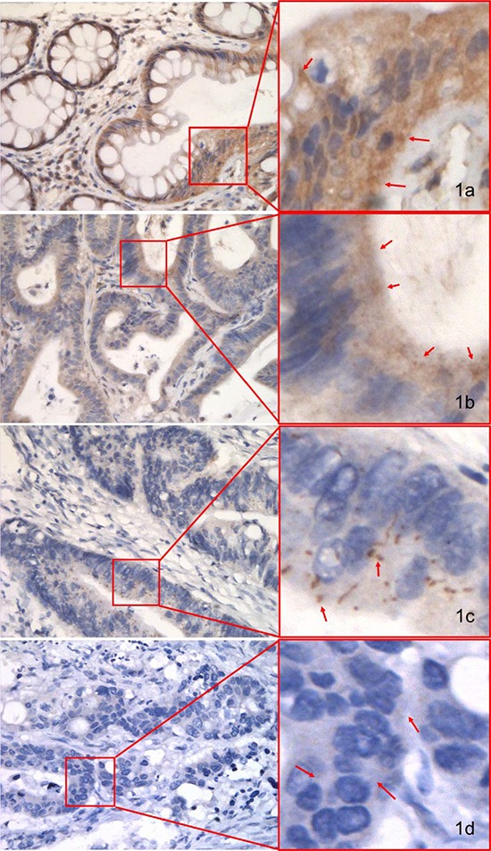
Figure 1. IHC staining of pMKK4 using selective immune-reactivity antibody with phosphopeptide containing S257/T261 site of MKK4 in normal colonic mucosa (NCM), colorectal adenoma (CA) and colorectal cancer (CRC) from the pilot set.

pMKK4 was cytoplasmic stained. Variable degree of pMKK4 expression level was showed in NCM (A, strong), CA (B, moderate) and CRC (C, weak) & (D, negative). Photos were captured by NIKKON microscope at 400×.
