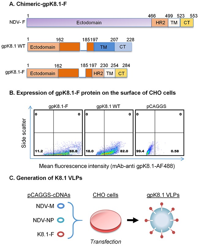
Figure 1. Schematic representation of the construction of chimeric KSHV gpK8.1-F and production of gpK8.1 VLPs.

A. gpK8.1-F plasmid constructs (not to scale) showing full-length NDV-F (top), full-length/wild-type (WT) gpK8.1 (middle), and chimeric gpK8.1-F (bottom). B. Flow cytometric analysis for surface expression of gpK8.1 protein on 106 CHO cells transfected with 1 μg of pCAGGS gpK8.1-F chimera, pCAGGS gpK8.1 WT (positive control), and empty pCAGGS vector (negative control) was performed at 48 h post-transfection. Transfected cells were resuspended in PBS, stained with anti-gpK8.1 mAb, which detects the ectodomain of the protein, followed by secondary antibody goat-anti-mouse IgG conjugated to AF488. From left, gpK8.1-F chimeric protein is robustly expressed on the surface of CHO cells, similar to WT gpK8.1 (middle panel), and no gpK8.1 protein was detected in CHO cells transfected with pCAGGS alone (negative control). C. Illustration of co-transfection of CHO cells with three cDNA plasmids required to make VLPs, pCAGGS cDNAs NDV-M, NDV-NP and gpK8.1-F, resulting in release of fully-assembled gpK8.1 VLPs. cDNAs NDV-M and NDV-NP have been described (see Materials and Methods).
